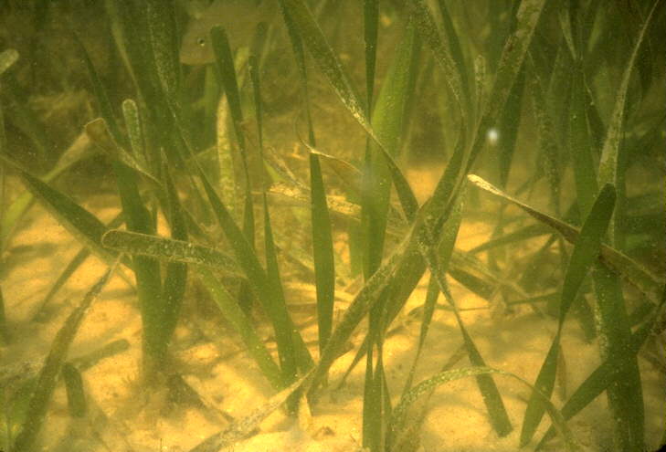
Figure 3. A Thalassia testudinum seagrass meadow where pinfish lurk. Photograph from St. Joe Bay, Florida (photo by Ron Phillips from the Univ. of Hawaii's Seagrass Home Page) |

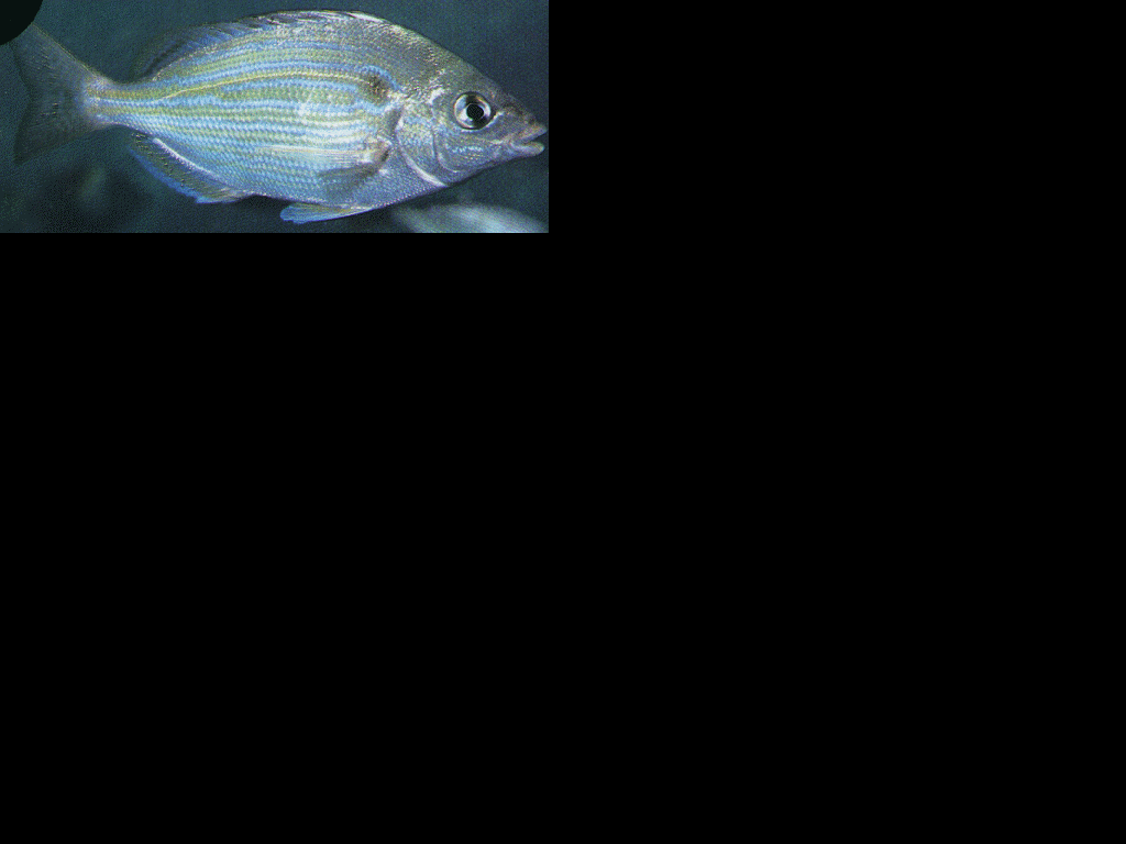
Figure 4. Seagrass (Zostera marina) inside a 120 mm SL pinfish stomach collecetd in Core Sound, NC. Some pinfish have > 90 % of their gut contents composed of seagrasses. Note the bite-sized pieces of the leaves. (photo by Joe Luczkovich) |
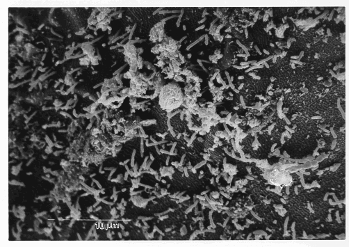
Figure 5. A scanning electron micrograph of rod-shaped cellulolytic symbiotic bacteria from pinfish intestines. (Photo by Tim Charles, ECU SEM laboratory) |